图书介绍
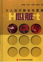
介入治疗解剖学图谱 心脏 中英文本2025|PDF|Epub|mobi|kindle电子书版本百度云盘下载

- 隋鸿锦,高连君,于胜波等主编 著
- 出版社: 沈阳:辽宁科学技术出版社
- ISBN:7538149821
- 出版时间:2007
- 标注页数:161页
- 文件大小:36MB
- 文件页数:181页
- 主题词:
PDF下载
下载说明
介入治疗解剖学图谱 心脏 中英文本PDF格式电子书版下载
下载的文件为RAR压缩包。需要使用解压软件进行解压得到PDF格式图书。建议使用BT下载工具Free Download Manager进行下载,简称FDM(免费,没有广告,支持多平台)。本站资源全部打包为BT种子。所以需要使用专业的BT下载软件进行下载。如BitComet qBittorrent uTorrent等BT下载工具。迅雷目前由于本站不是热门资源。不推荐使用!后期资源热门了。安装了迅雷也可以迅雷进行下载!
(文件页数 要大于 标注页数,上中下等多册电子书除外)
注意:本站所有压缩包均有解压码: 点击下载压缩包解压工具
图书目录
第一章 心脏介入治疗入路3
Ⅰ-1 大腿前内侧面的肌肉、血管和神经 Muscles,blood vessels and nerves of the anteromedial aspect of the thigh3
Ⅰ-2 腹股沟韧带中部横断面 Transverse section through the middle of inguinal ligament4
Ⅰ-3 股三角的血管和神经 Blood vessels and nerves of the femoral triangle5
Ⅰ-4 盆部和大腿上部的动脉(前面观) Arteries of pelvic cavity and the upper part of the thigh(anterior view)6
Ⅰ-5 盆部和大腿上部的动脉前面观(铸型标本) Arteries of pelvic cavity and the upper part of the thigh:anterior view(casting specimen)7
Ⅰ-6 盆部动脉造影(后前位) Injection of pelvic arteries in PA view8
Ⅰ-7 导管指示的髂血管走行 Iliac vein and artery guided by catheters9
Ⅰ-8 导管指示的下腔静脉和腹主动脉走行 Inferior caval vein and abdominal aorta by catheters9
Ⅰ-9 主动脉及其分支前面观(铸型标本) Aorta and its branches:anterior view(casting specimen)10
Ⅰ-10 上肢前面浅层的肌肉、血管和神经 Superficial Muscles,blood vessels and nerves of the anterior aspect of the upper limb11
Ⅰ-11 上肢浅层血管前内侧面观(铸型标本) Superficial Blood vessels of the upper limb:anterior-medial view (casting specimen)12
Ⅰ-12 上肢的动脉前内侧面观(铸型标本) Arteries of the upper limb:anterior-medial view(casting specimen)13
Ⅰ-13 臂部的动脉(前面观) Arteries of the upper arm(anterior view)14
Ⅰ-14 前臂的动脉(前面观) Arteries of the forearm(anterior view)14
Ⅰ-15 腋动脉及其分支 Axillary artery and its branches15
Ⅰ-16 腋窝的血管和神经 Blood vessels and nerves of the axilla16
Ⅰ-17 锁骨下窝 Infraclavicular fossa17
Ⅰ-18 锁骨下窝(胸大肌掀开) Infraclavicular fossa with pectoralis major folded17
Ⅰ-19 胸部经锁骨中线矢状断面 Thoracic sagittal section through the midclavicular line18
Ⅰ-20 胸部经胸锁关节横断面 Thoracic cross-section through the sternoclavicular joint18
Ⅰ-21 锁骨下静脉穿刺体表穿刺点 Subclavian vein puncture19
Ⅰ-22 X线透视下锁骨下静脉穿刺针走行 Subclavian vein puncture by X-ray19
Ⅰ-23 导丝指示的锁骨下静脉走行 Subclavian vein guided by wire20
Ⅰ-24 锁骨下动脉、腋动脉造影 Subclavian arteries and axillary artery angiography20
Ⅰ-25 颈部侧面观 Lateral view of the neck21
Ⅰ-26 颈部侧面观(示颈内静脉) Lateral view of the neck(showing internal jugular v.)22
Ⅰ-27 锁骨上小窝 Lesser supraclavicular fossa23
Ⅰ-28 颈部横断面 Cross-section of the neck24
Ⅰ-29 颈内静脉穿刺体表穿刺点 Internal jugular vein puncture24
Ⅰ-30 X线透视下颈内静脉穿刺针走行 Internal jugular vein puncture by X-ray25
Ⅰ-31 导丝指示的颈内静脉及上腔静脉走行 Internal jugular vein and superior vena cava guided by wire25
Ⅰ-32 左上腔静脉畸形(导丝显示) Abnormal dranage from left superior vena cava demonstrated by wire26
Ⅰ-33 左上腔静脉畸形(造影显示) Abnormal dranage from left superior vena cava demonstrated by angiography26
第二章 心律失常的介入治疗29
Ⅱ-1 心脏传导系 Conducting system of the heart29
Ⅱ-2 心脏电生理检查常规放置导管及血管入路(后前位) Routine catheters position of cardial electriophysiological study(PA)30
Ⅱ-3 心脏电生理检查常规放置导管及血管入路(左前斜位45°) Routine catheters position of cardial electriophy siological study(LAO)30
第一节 房室结双径路的导管射频消融 Catheter ablation of duai atrioventricular node reentry tachycardia31
Ⅱ-4 右心房内腔右上面观 Superior right view of interior of right atrium31
Ⅱ-5 房间隔和室间隔右侧面观 Right lateral view of interatrial and interventricular septums32
Ⅱ-6 右心房内腔右后面观 Posterior right view of interior of right atrium33
Ⅱ-7 房室结双径路的慢径区导管消融(A)(右前斜位30°) Slow pathway catheter ablation of dual atrioventricular node reentry tachycardia(A)(RAO30°)34
Ⅱ-8 房室结双径路的慢径区导管消融(B)(右前斜位30°) Slow pathway catheter ablation of dual atrioventricular node reentry tachycardia(B)(RAO30°)34
第二节 左侧房室旁路的导管射频消融 Catheter ablation of left atrioventricle accessory pathway35
Ⅱ-9 心室底(示房室口) Base of the ventricles(showing atrioventricular orifices)35
Ⅱ-10 左心室内腔(左侧面观) The lateral view of interior of left atrium36
Ⅱ-11 左后壁旁路导管消融(右前斜位30°) Left posterior free wall accessory pathway catheter ablation(RAO 30°)37
Ⅱ-12 左游离壁旁路导管消融(右前斜位30°) Left free wall accessory pathway catheter ablation(RAO 30°)37
Ⅱ-13 左游离壁旁路导管消融(左前斜位45°) Left free wall accessory pathway catheter ablation(LAO 45°)38
Ⅱ-14 左游离壁旁路导管消融(后前位) Left free wall accessory pathway catheter ablation(PA)38
Ⅱ-15 左后间隔旁路导管消融(右前斜位30°) Left posterior septal accessory pathway catheter ablation(RAO 30°)39
Ⅱ-16 左后间隔旁路导管消融(左前斜位45°) Left posterior septal accessory pathway catheter ablation(LAO 45°)39
Ⅱ-17 左后间隔旁路导管消融(后前位) Left posterior septal accessory pathway catheter ablation(PA)40
Ⅱ-18 主动脉逆行途径记录到的HIS电位区域(右前斜位30°) HIS bundle potential recorded at left septum(RAO 30°)40
Ⅱ-19 主动脉逆行途径记录到的HIS电位区域(左前斜位45°) HIS bundle potential recorded at left septum(LAO 45°)41
Ⅱ-20 主动脉逆行途径记录到的HIS电位区域(后前位) HIS bundle potential recorded in left septum(PA)41
第三节 右侧房室旁路的导管射频消融 Catheter ablation of right atrioventricle accessory pathway42
Ⅱ-21 左心房内腔(示右肺静脉口) Interior of left atrium(showing right orifices of pulinonary veins)42
Ⅱ-22 心室底(示左、右房室口) Base of the ventricles(showing right and left atrioventricular orifices)42
Ⅱ-23 右心室内腔 Interior of right ventricle43
Ⅱ-24 右游离壁旁路导管消融(左前斜位45°) Right free wall accessory pathway catheter ablation(LAO 45°)44
Ⅱ-25 右后壁旁路导管消融(左前斜位45°) Right posterior accessory pathway catheter ablation(LAO 45°)44
第四节 心房扑动的导管射频消融 Catheter ablation of atrial flutter45
Ⅱ-26 右心房内腔 Interior of right atrium45
Ⅱ-27 右心房内腔右上面观 Superior right view of interior of right atrium45
Ⅱ-28 右心房打开右上面观 Superior right view of opened right atrium46
Ⅱ-29 典型心房扑动导管消融(左前斜位45°) Linear ablation of typical atrial flutter catheter(LAO 45°)46
第五节 室早、室速的导管射频消融 Catheter ablation of ventricular arrhythmia47
Ⅱ-30 右心室和肺动脉干打开前面观 Anterior view of opened right ventricle and pulmonary trunk47
Ⅱ-31 房室口和动脉口前面观(心室切除) Anterior view of atrioventricular orifices,orifice of pulmonary trunk and aortic orifice(ventricles removed)48
Ⅱ-32 右心室内腔前面观 Anterior view of interior of right ventricle49
Ⅱ-33 右室流出道 Outflow tract of right ventricle50
Ⅱ-34 右房室口、肺动脉口和右房前面观(心室切除) Anterior view of right atrioventricular orifices,orifice of pulmonary trunk and right atrium(ventricles removed)50
Ⅱ-35 右室流出道室速导管消融(后前位) Right ventricular outflow tract tachycardia catheter ablation(PA)51
Ⅱ-36 右室流出道室速导管消融(左前斜位45°) Right ventricular outflow tract tachycardia catheter ablation(LAO45°)51
Ⅱ-37 右室流出道室速导管消融(右前斜位30°) Right ventricular outflow tract tachycardia catheter ablation(RAO30°)52
Ⅱ-38 心室底(示主动脉口) Base of the ventricles(showing aortic orifice)52
Ⅱ-39 左心室内腔(沿长轴观) Interior of left atrium(the view along the long axis)53
Ⅱ-40 左心室内腔(左侧面观) The lateral view of interior of left atrium53
Ⅱ-41 左心室打开左前面观 Left anterior view of opened left ventricle54
Ⅱ-42 房室口和动脉口前面观(心室切除) Anterior view of atrioventricular orifices,orifice of pulmonary trunk and aortic orifice (ventricles removed)54
Ⅱ-43 特发性左室间隔部室速导管消融(后前位) Idiopathic left ventricle septal tachycardia catheter ablation(PA)55
Ⅱ-44 特发性左室间隔部室速导管消融(右前斜位30°) Idiopathic left ventricle septal tachycardia catheter ablation(RAO 30°)55
Ⅱ-45 特发性左室间隔部室速导管消融(左前斜位45°) Idiopathic left ventricle septal tachycardia catheter ablation(LAO 45°)55
Ⅱ-46 左室流出道室速导管消融(主动脉窦内) Left ventricular outflow tract tachycardia catheter ablation(interior aortic sinus)56
Ⅱ-47 左室流出道室速导管消融(主动脉瓣下) Left ventricular outflow tract tachycardia catheter ablation(below the aortic valve)56
第六节 心房颤动的导管射频消融 Catheter ablation of atrial fibrillation57
Ⅱ-48 左、右心室和右心房剖面 Sections of left,right ventricles and right atrium57
Ⅱ-49 右心房内腔 Interior of right atrium58
Ⅱ-50 右心房内腔右上面观 Superior right view of interior of right atrium58
Ⅱ-51 卵圆窝(经右房室口) Fossa ovalis(through the right atrioventricular orifice)59
Ⅱ-52 右心房下面观(下腔静脉口打开,房壁上翻) Inferior view of right atrium with orifice of inferior vena cava opened and wall of atrium reflected upwards59
Ⅱ-53 房间隔穿刺(后前位) Interatrial septum puncture(PA)60
Ⅱ-54 房间隔穿刺(右前斜位45°) Interatrial septum puncture(RAO 45°)60
Ⅱ-55 左心房内腔(示右肺静脉口) Interior of left atrium(showing right orifices of pulmonary veins)61
Ⅱ-56 心脏、气管和食管右侧面观 Anterior view of heart,trachea and esophagus62
Ⅱ-57 左、右心房剖面(下肺静脉口) Section of left and right atria(orifices of inferior pulmonary veins)63
Ⅱ-58 左、右心房剖面(上肺静脉口) Section of left and right atria(orifices of superior pulmonary veins)64
Ⅱ-59 左心房打开右后面观 Posterior right view of opened left atrium65
Ⅱ-60 左、右心房剖面(右肺静脉口) Section of left and right atria(orifices of right pulmonary veins)66
Ⅱ-61 左心房打开右后上面观 Superior posterior right view of opened left atrium67
Ⅱ-62 心脏、气管和食管后面观 Posterior view of heart,trachea and esophagus68
Ⅱ-63 纵隔右侧面观 Right lateral view of mediastinum69
Ⅱ-64 心脏左前面观(Marshall韧带) The anterior left surface of the heart(ligament of Marshall)70
Ⅱ-65 右心房打开下面观 Inferior view of opened right atrium71
Ⅱ-66 左上肺静脉逆行造影(左前斜位45°) Retrograde angiography of left superior pulmonary vein(LAO45°)72
Ⅱ-67 右上肺静脉逆行造影(A)(左前斜位45°) Retrograde angiography of right superior pulmonary vein(A)(LAO45°)72
Ⅱ-68 右上肺静脉逆行造影(B)(左前斜位45°) Retrograde angiography of right superior pulmonary vein(B)(LAO45°)73
Ⅱ-69 肺静脉节段性电隔离(左上肺静脉)(左前斜位) Segmental pulmonary vein isolation(left superior pulmonary vein)(LAO)73
Ⅱ-70 左心耳与左上肺静脉位置影像图(右前斜位45°) Left atrium and left superior pulmonary vein angiography(RAO45°)74
Ⅱ-71 左心耳与左上肺静脉位置影像图(左前斜位45°) Left atrium and left superior pulmonary vein angiography(LAO45°)74
Ⅱ-72 肺静脉节段性电隔离(左下肺静脉)(左前斜位) Segmental pulmonary vein isolation(left inferior pulmonary vein)(LAO)75
Ⅱ-73 肺静脉节段性电隔离(右上肺静脉)(左前斜位) Segmental pulmonary vein isolation(right superior pulmonary vein)(LAO)75
Ⅱ-74 肺静脉节段性电隔离(右下肺静脉)(左前斜位) Segmental pulmonary vein isolation(right inferior pulmonary vein)(LAO)75
Ⅱ-75 多层螺旋CT正常肺静脉成像 Normal pulmonary vein imaging by multiple slice spiral CT76
Ⅱ-76 多层螺旋CT肺静脉成像(肺静脉狭窄) Pulmonary vein stenosis imaging by multiple slice spiral CT76
Ⅱ-77 心脏铸型(后面观) Casting specimen of heart(posterior view)77
Ⅱ-78 心脏后面观 Posterior view of heart78
第七节 起搏器植入 Pacemaker inplantation79
Ⅱ-79 右心室内腔 Interior of right ventricle79
Ⅱ-80 右心室打开前面观 Anterior view of opened right ventricle79
Ⅱ-81 心室单腔起搏(后前位) Single chamber pacing(PA)80
Ⅱ-82 心室单腔起搏(右前斜位) Single chamber pacing(RAO)80
Ⅱ-83 右心房内腔 Interior of right atrium81
Ⅱ-84 双腔起搏(后前位) Dual chamber pacing(PA)82
Ⅱ-85 双腔起搏(右前斜位) Dual chamber pacing(RAO)82
Ⅱ-86 左心室后外侧壁(心尖下垂) The posterior lateral wall of left atrium(cardiac apex downward)83
Ⅱ-87 冠状窦铸型(下面观) Inferior view of coronary sinus(casting specimen)83
Ⅱ-88 心脏血管铸型(心室底,示冠状窦) The casting specimen of the vessels of the heart(base of the ventricles,showing coronary sinus)84
Ⅱ-89 心脏血管铸型下面观(A) Inferior view of the vessels of the heart(casting specimen)85
Ⅱ-90 心脏血管铸型下面观(B) Inferior view of the vessels of the heart(casting specimen)86
Ⅱ-91 心脏血管铸型下面观(C) Inferior view of the vessels of the heart(casting specimen)87
Ⅱ-92 冠状静脉窦逆行造影 Retrograde angiography of coronary sinus88
Ⅱ-93 双心室起搏(后前位) Dual ventricle pacing(PA)88
Ⅱ-94 双心室起搏(右前斜位) Dual ventricle pacing(RAO)89
Ⅱ-95 双心室起搏(左前斜位) Dual ventricle pacing(LAO)89
Ⅱ-96 单腔埋藏式自动复律除颤器(ICD)(后前位) Single chamber automatic implanted cardioversion defibrillator(PA)90
Ⅱ-97 单腔埋藏式自动复律除颤器(ICD)(右前斜位) Single chamber automatic implanted cardioversion defibrillator(RAO)90
Ⅱ-98 双腔埋藏式自动复律除颤器(ICD)(后前位) Dual chamber automatic implanted cardioversion defibrillator(PA)91
Ⅱ-99 双腔埋藏式自动复律除颤器(ICD)(右前斜位) Dual chamber automatic implanted cardioversion defibrillator(RAO)91
Ⅱ-100 双腔埋藏式自动复律除颤器(ICD)(左前斜位) Dual chamber automatic implanted cardioversion defibrillator(LAO)92
第八节 心包填塞 Cardiac tamponade93
Ⅱ-101 心包和肺前面观 Anterior view of pericardium and lung93
Ⅱ-102 心脏前面观(心包打开) Anterior view of heart with opened pericardial sac94
Ⅱ-103 心脏前面观(心包打开,肺摘除) Anterior view of heart with opened pericardial sac and lunges removed95
Ⅱ-104 心包腔(心脏摘除)前面观 Pericardial sac with heart removed,anterior view96
Ⅱ-105 心包填塞(A)(左前斜位45°) Cardiac tamponade(A)(LAO 45°)97
Ⅱ-106 心包填塞(B)(左前斜位45°) Cardiac tamponade(B)(LAO 45°)97
Ⅱ-107 心包填塞(C)(左前斜位45°) Cardiac tamponade(C)(LAO 45°)98
第三章 冠心病的介入治疗101
Ⅲ-1 心脏右前面观 The anterior right surface of the heart101
Ⅲ-2 心脏右侧面观 The right surface of the heart102
Ⅲ-3 心脏左前面观 The anterior left surface of the heart103
Ⅲ-4 心脏左侧面观 The left surface of the heart104
Ⅲ-5 心脏前面观(示冠脉) The anterior surface of the heart(showing coronary arteries)105
Ⅲ-6 心脏左前上面观(示冠脉) The anterior superior left surface of the heart(showing coronary arteries)106
Ⅲ-7 心脏膈面观(示冠脉) The diaphragmatic surface of the heart(showing coronary arteries)107
Ⅲ-8 心脏右前上面观(示冠脉) The anterior right surface of the heart(showing coronary arteries)108
Ⅲ-9 左心室打开前面观 Anterior view of opened left ventricle109
Ⅲ-10 左心室打开左侧面观 Left lateral view of opened left ventricle110
Ⅲ-11 心脏血管铸型前面观 Anterior view of the vessels of the heart(casting specimen)111
Ⅲ-12 心脏血管铸型右前面观 Right anterior view of the vessels of the heart(casting specimen)112
Ⅲ-13 心脏血管铸型右后面观 Right posterior view of the vessels of the heart(casting specimen)113
Ⅲ-14 心脏血管铸型膈面观 Diaphragmatic aspect of the vessels of the heart(casting specimen)114
Ⅲ-15 心脏血管铸型左前面观(A) Left anterior view of the vessels of the heart(casting specimen)115
Ⅲ-16 心脏血管铸型左前面观(B) Left anterior view of the vessels of the heart(casting specimen)116
Ⅲ-17 心脏冠状动脉前面观(A) Anterior view of the coronary arteries of the heart(casting specimen)117
Ⅲ-18 心脏冠状动脉前面观(B) Anterior view of the coronary arteries of the heart(casting specimen)118
Ⅲ-19 心脏冠状动脉前面观(C) Anterior view of the coronary arteries of the heart(casting specimen)119
Ⅲ-20 心脏冠状动脉铸型下面观(A) Inferior view of the coronary arteries of the heart(casting specimen)120
Ⅲ-21 心脏冠状动脉铸型下面观(B) Inferior view of the coronary arteries of the heart(casting specimen)121
Ⅲ-22 心脏冠状动脉铸型下面观(C)Inferior view of the coronary arteries of the heart(casting specimen)121
Ⅲ-23 左冠状动脉铸型标本镜像(A)(左前斜位,足位) Mirror image of casting specimen of left coronary artery(LAO,caudal)122
Ⅲ-24 左冠状动脉铸型标本镜像(B)(左前斜位,足位) Mirror image of casting specimen of left coronary artery(LAO,caudal)122
Ⅲ-25 左冠状动脉铸型标本镜像(C)(左前斜位,足位) Mirror image of casting specimen of left coronary artery(LAO,caudal)123
Ⅲ-26 正常左冠状动脉造影(左前斜位50°,足位30°) Normal left coronary artery angiography(LAO 50°,caudal 30°)123
Ⅲ-27 左冠状动脉铸型标本(A)(右前斜位,足位) Casting specimen of left coronary artery(RAO,caudal)124
Ⅲ-28 左冠状动脉铸型标本(A)(镜像) Mirror image of casting specimen of left coronary artery124
Ⅲ-29 左冠状动脉铸型标本(B)(镜像)(右前斜位,足位) Mirror image of casting specimen of left coronary artery(RAO,caudal)125
Ⅲ-30 正常左冠状动脉造影(右前斜位30°,足位30°) Normal left coronary artery angiography(RAO 30°,caudal 30°)125
Ⅲ-31 左冠状动脉铸型标本(A)(右前斜位,头位) Casting specimen of left coronary artery(RAO,cranial)126
Ⅲ-32 左冠状动脉铸型标本(B)(右前斜位,头位) Casting specimen of left coronary artery(RAO,cranial)127
Ⅲ-33 正常左冠状动脉造影(右前斜位30°,头位30°) Normal left coronary artery angiography(RAO 30°,cranial 30°)128
Ⅲ-34 左冠状动脉铸型标本(A)(左前斜位,头位) Casting specimen of left coronary artery(LAO,cranial)129
Ⅲ-35 左冠状动脉铸型标本(B)(左前斜位,头位) Casting specimen of left coronary artery(LAO,cranial)129
Ⅲ-36 正常左冠状动脉造影(左前斜位50°,头位30°) Normal left coronary artery angiography(LAO 50°,cranial 30°)130
Ⅲ-37 右冠状动脉铸型标本(A)(左前斜位) Casting specimen of right coronary artery(LAO)131
Ⅲ-38 右冠状动脉铸型标本(B)(左前斜位) Casting specimen of right coronary artery(LAO)132
Ⅲ-39 正常右冠状动脉造影(左前斜位50°) Normal right coronary artery angiography(LAO 50°)133
Ⅲ-40 右冠状动脉铸型标本(A)(左前斜位,头位) Casting specimen of right coronary artery(LAO,cranial)134
Ⅲ-41 右冠状动脉铸型标本(B)镜像(左前斜位,头位) Mirror image of casting specimen of right coronary artery(LAO,cranial)135
Ⅲ-42 正常右冠状动脉造影(左前斜位50°,头位30°) Normal right coronary artery angiography(LAO 50°,cranial 30°)136
Ⅲ-43 右冠状动脉铸型标本(A)(右前斜位) Casting specimen of right coronary artery(RAO)137
Ⅲ-44 右冠状动脉铸型标本(B)(右前斜位) Casting specimen of right coronary artery(RAO)138
Ⅲ-45 正常右冠状动脉造影(右前斜位30°) Normal right coronary artery angiography(RAO 30°)139
Ⅲ-46 左心室造影(舒张期)(右前斜位30°) Left ventricle angiography(diastolic period)(RAO 30°)140
Ⅲ-47 左心室造影(收缩期)(右前斜位30°) Left ventricle angiography(systolic period)(RAO 30°)140
Ⅲ-48 左心室和升主动脉(打开)左前面观 Left anterior view of opened left ventricle and ascending aorta141
Ⅲ-49 主动脉根部造影(左前斜位30°) Aortic root angiography(LAO 30°)142
Ⅲ-50 多层螺旋CT冠状动脉成像(A) Coronary artery imaging by multiplayer spiral CT(A)142
Ⅲ-51 多层螺旋CT冠状动脉成像(B) Coronary artery imaging by multiplayer spiral CT(B)143
Ⅲ-52 病例一:前降支中段狭窄(头位) Case one:stenosis in middle of left anterior descending coronary artery(cranial)143
Ⅲ-53 病例一:前降支中段狭窄(右前斜位,头位) Case one:stenosis in middle of left anterior descending coronary artery(RAO,cranial)144
Ⅲ-54 病例二:前降支开口狭窄(左前斜位,足位) Case two:stenosis in ostium of left anterior descending coronary artery(LAO,caudal)144
Ⅲ-55 病例二:前降支开口狭窄(右前斜位,足位) Case two:stenosis in ostium of left anterior descending coronary artery(RAO,caudal)145
Ⅲ-56 病例二:前降支开口狭窄(足位) Case two:stenosis in ostium of left anterior descending coronary artery(caudal)145
Ⅲ-57 病例三:前降支中段狭窄(头位) Case three:stenosis in middle of left anterior descending coronary artery(cranial)146
Ⅲ-58 病例四:前降支中段肌桥(舒张期) Case four:myocardial bridging in middle of left anterior descending coronary artery(diastolic period)146
Ⅲ-59 病例四:前降支中段肌桥(收缩期) Case four:myocardial bridging in middle of left anterior descending coronary artery(systolic period)147
Ⅲ-60 心脏左前面观(冠状动脉和肌桥) The anterior left surface of the heart(coronary artery and myocardial bridging)147
Ⅲ-61 病例五:前降支近段完全闭塞(左前斜位,足位) Case five:total occlusion proximal left anterior descending coronary artery(LAO,caudal)148
Ⅲ-62 病例五:前降支近段完全闭塞(足位) Case five:total occlusion proximal left anterior descending coronary artery(caudal)148
Ⅲ-63 病例五:前降支近段完全闭塞(头位) Case five:total occlusion proximal left anterior descending coronary artery(cranial)149
Ⅲ-64 病例五:前降支近段完全闭塞(左前斜位,头位) Case five:total occlusion proximal left anterior descending coronary artery(LAO,cranial)149
Ⅲ-65 病例六:回旋支中段完全闭塞(右前斜位,足位) Case six:total occlusion in middle of left circumflex artery150
Ⅲ-66 病例七:右冠状动脉中段高度狭窄(左前斜位) Case seven:serious stenosis in middle of right coronary artery(LAO)150
Ⅲ-67 病例八:右冠状动脉近段完全闭塞(左前斜位) Case eight:total occlusion proximal right coronary artery(LAO)151
Ⅲ-68 病例九:右冠状动脉中段闭塞病变治疗前 Case nine:total occlusion in middle of right coronary artery151
Ⅲ-69 病例九:右冠状动脉中段闭塞病变球囊扩张 Case nine:balloon dilation in middle of right coronary artery152
Ⅲ-70 病例九:右冠状动脉中段闭塞病变支架植入后 Case nine:right coronary artery after stent implantation152
第四章 先天性心脏病介入治疗155
Ⅳ-1 房间隔和室间隔(隔侧瓣切除)右侧面观 Right lateral view of interatrial and interventricular septums with septal cusp removed155
Ⅳ-2 房间隔缺损经封堵器治疗后(后前位) Atrial septal defect treated by occluder(PA)156
Ⅳ-3 房间隔缺损经封堵器治疗后(左前斜位) Atrial septal defect treated by occluder(LAO)156
Ⅳ-4 心脏水平切:心房、心室和室间隔 Horizontal section of heart:atria,ventricles and interventricular septum157
Ⅳ-5 室间隔缺损左室造影(左前斜位,头位) Left ventricle angiography of ventricular septal defect(LAO,cranial)158
Ⅳ-6 室间隔缺损封堵器植入后左室造影(左前斜位,头位) Left ventricle angiography of ventricular septal defect after occluder implantation(LAO,cranial)158
Ⅳ-7 室间隔缺损经封堵器治疗后(后前位) Ventricular septal defect treated by occluder(PA)158
Ⅳ-8 心脏前上面观(动脉韧带) Superior anterior surface of the heart(arterial ligament)159
Ⅳ-9 动脉导管未闭主动脉造影(左侧位) Aortic angiography of patent ductus arteriosus(left lateral projection)160
Ⅳ-10 动脉导管未闭封堵器植入后主动脉造影(左侧位) Aortic angiography of patent ductus arteriosus after occluder implantation(left lateral projection)160
Ⅳ-11 动脉导管未闭经封堵器治疗后(左侧位) Patent ductus arteriosus treated by occluder(left lateral projection)161
Ⅳ-12 动脉导管未闭经封堵器治疗后(后前位) Patent ductus arteriosus treated by occluder(PA)161
热门推荐
- 1167284.html
- 3339530.html
- 2044169.html
- 3671007.html
- 1122862.html
- 2909186.html
- 250979.html
- 1740954.html
- 661454.html
- 2131895.html
- http://www.ickdjs.cc/book_1528560.html
- http://www.ickdjs.cc/book_946299.html
- http://www.ickdjs.cc/book_623278.html
- http://www.ickdjs.cc/book_2359854.html
- http://www.ickdjs.cc/book_3189170.html
- http://www.ickdjs.cc/book_1617855.html
- http://www.ickdjs.cc/book_2788415.html
- http://www.ickdjs.cc/book_2594724.html
- http://www.ickdjs.cc/book_3503913.html
- http://www.ickdjs.cc/book_2678837.html